Identification of cell-autonomous microglia phenotypes in an induced pluripotent stem cell model of Huntington’s disease
Abstract
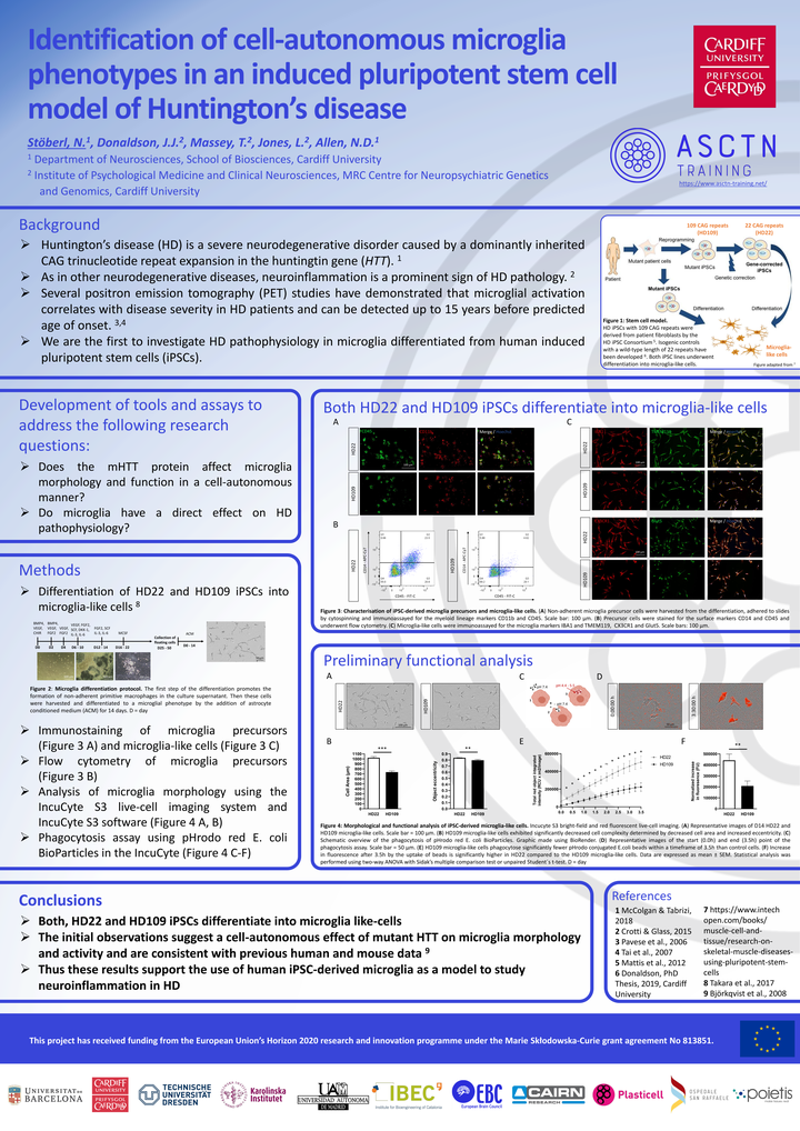
Huntington’s disease (HD) is a severe neurodegenerative disorder caused by a dominantly inherited CAG trinucleotide repeat expansion in the huntingtin gene (HTT). As in other neurodegenerative diseases, neuroinflammation is a prominent sign of HD pathology. Microglia are the principal resident innate immune cells of the CNS. Several positron emission tomography (PET) studies have demonstrated that microglial activation correlates with disease severity and is evident in presymptomatic HD patients up to 15 years before predicted age of onset. Nonetheless, an open question is whether mutant HTT expression leads to cell-autonomous phenotypes in microglia, which potentially contribute to disease onset and progression. To address this, we used patient-derived induced pluripotent stem cell (iPSC) models of HD microglia with 109 CAG repeats (HD109) and isogenic controls with a corrected wild-type length of 22 repeats (HD22). Differentiated iPSC-derived myeloid precursors and microglia-like cells were characterized by flow cytometry and immunohistochemistry for the expression of cell-specific markers. Both HD109 and HD22 isogenic control cells express desired microglia markers. We found that HD109 microglia exhibit significantly decreased cell complexity determined by cell area and eccentricity. The phagocytosis of pathogens, apoptotic cells and debris is a key feature of microglia function. Phagocytosis of E.coli beads was significantly decreased in HD109 microglia compared to HD22 isogenic controls. These initial observations suggest a cell-autonomous effect of mutant HTT on microglia status and activity and are consistent with previous data on microglial activation, thus supporting the use of iPSC-derived microglia as a model to study neuroinflammation in HD.